A tool to see the invisible in a wound
The discovery of a new tool to visualize the tissue which responds to an injury could revolutionize wound research and open new avenues in clinical diagnosis. Ring and Dworak et al., from our biomedical research group lead by Mikolaj Ogrodnik, have discovered a powerful new wound healing marker, coined the phosphorylated ribosomal protein S6 (p-rpS6) zone.
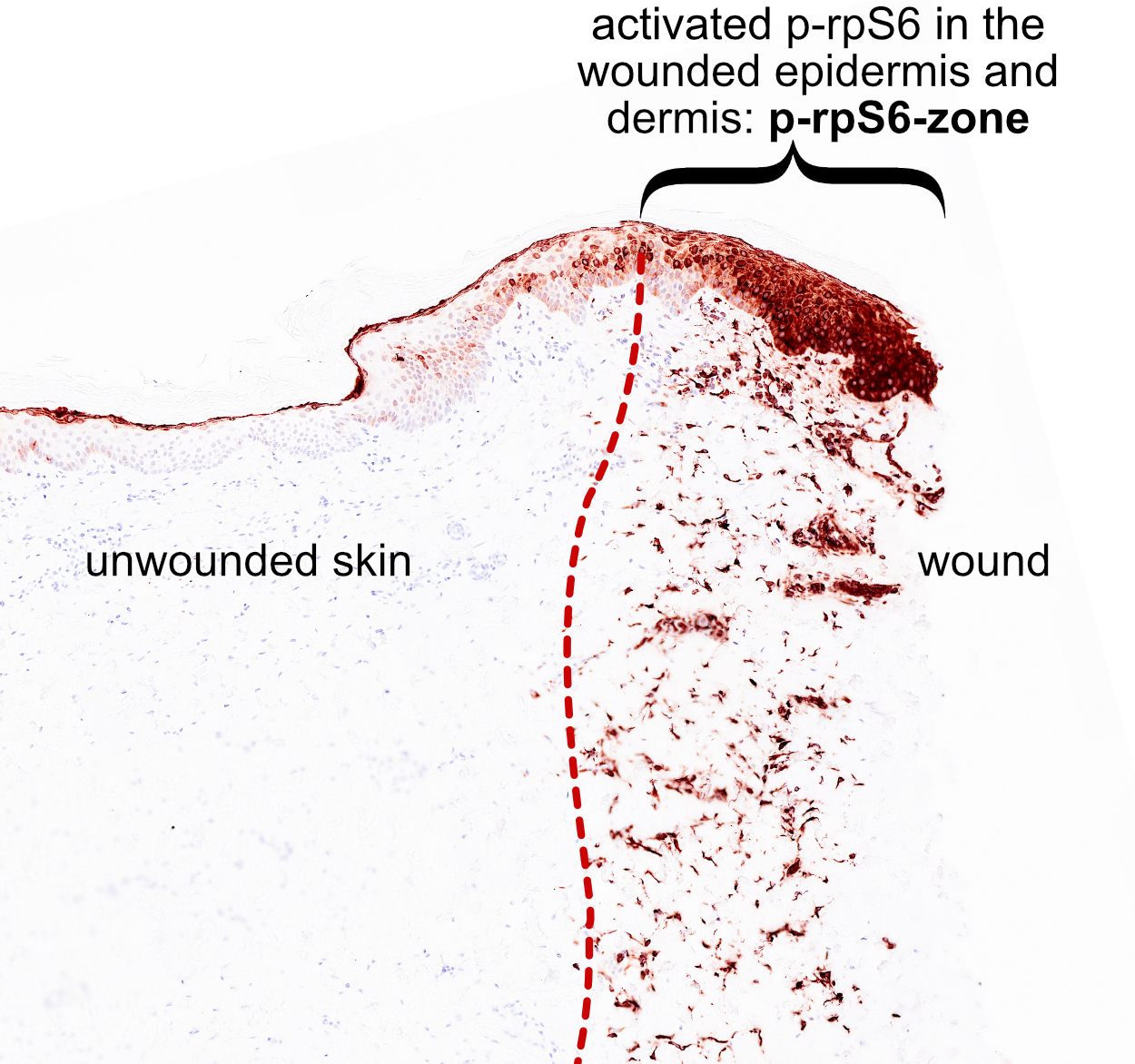
Using histology, the p-rpS6-zone highlights wounded skin tissue that is actively involved in the healing process, visually separating it from the wounded, dying tissue, as well as from the unaffected healthy tissue beyond the wound. The p-rpS6-zone is a marker of injured tissue detectable within minutes after injury and persists throughout the whole healing process. The p-rpS6-zone is an important discovery that unveils the “where” of wound healing and opens the doors to potential diagnostic and therapeutic applications. The research team’s findings have been published with Developmental Cell: The p-rpS6-zone delineates wounding responses and the healing process: Developmental Cell.
The zone as a tool. A skin injury causes cell death and bleeding, to which the injured tissue responds with blood clotting, blood vessel constriction, and a mobilization of immune cells. Finally, the tissue can regenerate the damaged tissue structures involving many simultaneous processes: increased cell division, blood vessel development, and an induction of cellular senescence. While the timing and sequence of wound healing processes is well understood, little is known about where exactly these processes are taking place within the tissue. This is because the signals that inform the tissue of an injury are both rapid and transient, happening within seconds and disappearing soon after, and thus are usually invisible to researchers without the help of costly and specialized tools. Until now. Ogrodnik and his team have shown that the p-rpS6-zone can be used to visualize the immediate spatial tissue response to wounding, highlighting the parts of the tissue which are responding to the wound and actively participating in wound healing, but are not damaged themselves.
What exactly is p-rpS6? The ribosomal protein S6 is an integral component of the machinery responsible for producing all proteins in a cell. Like other proteins, its surface can be modified by a process called phosphorylation, hence p-rpS6. In this case, this modification is associated with protein production and cell growth. While the skin normally has only a very small amount of phosphorylated rpS6, this situation changes as soon as any kind of wounding damages the skin, be it a burn, a cut, or a simple small prick of a needle. The p-rpS6-zone is then visible within minutes and until the wound is completely healed.
Features of the zone. The p-rpS6-zone is stable in time and space, and easy to detect. “Just to illustrate its reliability, researchers usually have to use high-magnification microscopes to detect damage caused to skin because of an injury, while the p-rpS6-zone can be seen with the naked eye on a stained histology sample,” says Mikolaj Ogrodnik. A further feature of the zone is that it encompasses all major skin cell types – fibroblasts, keratinocytes, endothelial cells – and encompasses all the major cellular processes of healing: increased cell division, cell enlargement, induction of cellular senescence, and blood vessel growth.
Where does the zone come from and what does it do? The p-rpS6-zone is activated by small damage-associated molecules (DAMPs) released by injured cells, and is dependent on oxygen availability. This makes it an interesting tool to study skin wounds in which a lack of blood supply and oxygen are an issue. As for the function of p-rpS6 itself, the team used a transgenic mouse model unable to phosphorylate this protein, and found that while healing still occurred, the overall process was slowed, resulting in increased scarring and decreased blood vessel formation.
The results generated by Ring and Dworak et al. from the Ogrodnik group have shown how healing properties are distributed around the wound. The next step is to develop the p-rpS6-zone as a clinical tool for medical diagnosis and an evaluation of the healing process.
Source: Ring, N. A. R., Dworak, H., Bachmann, B., Schaedl, B., Valdivieso, K., Rozmaric, T., … & Ogrodnik, M. (2023). The p-rpS6-zone delineates wounding response and the healing process. Developmental Cell. https://doi.org/10.1016/j.devcel.2023.04.001